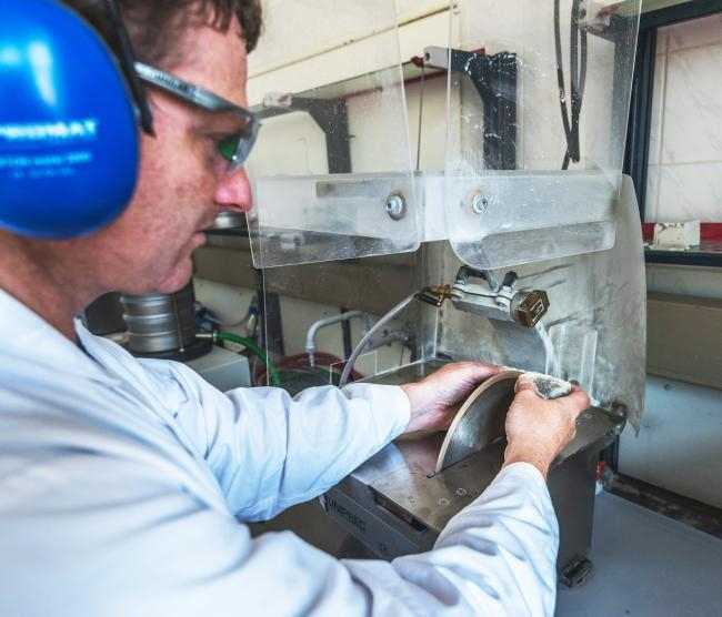
The Geo Laboratory is equipped for a wide range of studies in marine sciences. Thin sections and SEM specimens can be prepared from a variety of geological and biological sample material for detailed ultra-structural studies and then analysed using the light microscope and SEM. The SEM is equipped with different detectors to examine material properties.
The facilities offer the possibility to investigate the composition of marine samples with a focus on biotic carbonate components (e.g. calcareous algae, bryozoans, echinoderms and molluscs). Moreover, the external and internal structure of individual skeletal components can be analyzed using X-ray microtomography techniques.
Technical assistance is provided for sample pre-treatment and preparation, like carbon coating or gold sputtering, as well as for analytical methods and data interpretation.
| Laboratory equipment | Application |
| Stone saw, linear precision saw and high-precision grinding machines | porosity and material staining in thin sections |
| Sieving machine (Retsch LS200) for wet and dry sieving | particle size analyses in the range of 63 μm to 2 mm |
| Laser particle sizer (Horriba LA950) | particle size analyses in the range of 0.01 µm to 3 mm |
| Petrographic polarised light microscope (Leica DM-EP) with video support | thin section analysis |
| Cressington Coater (carbon and gold coating) | SEM sample preparation |
| Peltier cooling stage | wet or cool SEM sample investigations |
| SEM (Tescan Vega 3 XMU) with secondary electron detector (SE) | ultra-structural studies |
| Additional SEM equipment | |
| Backscatter electron diffraction detector (BSED) | material composition |
| Oxford energy-dispersive X-ray detector (EDX) | element composition |
| Low-vacuum mode | delicate or wet sample analyses |
| Skyscan Micro-CT for SEM | internal microstructure of samples <4 mm |


